Cyanobacteria
| Cyanobacteria | |
|---|---|
 |
|
| Scientific classification | |
| Domain: | Bacteria |
| Phylum: | Cyanobacteria |
| Orders | |
|
The taxonomy is currently under revision.[1] |
|

Cyanobacteria (also known as blue-green algae, blue-green bacteria, and Cyanophyta) is a phylum of bacteria that obtain their energy through photosynthesis. The name "cyanobacteria" comes from the color of the bacteria (Greek: κυανός (kyanós) = blue).
The ability of cyanobacteria to perform oxygenic photosynthesis is thought to have converted the early reducing atmosphere into an oxidizing one, which dramatically changed the composition of life forms on Earth by stimulating biodiversity and leading to the near-extinction of oxygen-intolerant organisms. According to endosymbiotic theory, chloroplasts in plants and eukaryotic algae have evolved from cyanobacteria via endosymbiosis.
Contents |
Ecology
Cyanobacteria can be found in almost every conceivable environment, from oceans to fresh water to bare rock to soil. They can occur as planktonic cells or form phototrophic biofilms in fresh water and marine environments, they occur in damp soil, or even temporarily moistened rocks in deserts. A few are endosymbionts in lichens, plants, various protists, or sponges and provide energy for the host. Some live in the fur of sloths, providing a form of camouflage. Aquatic cyanobacteria are probably best known for the extensive and highly visible blooms that can form in both freshwater and the marine environment and can have the appearance of blue-green paint or scum. The association of toxicity with such blooms has frequently led to the closure of recreational waters when blooms are observed. Marine bacteriophage are a significant parasite of unicellular, marine cyanobacteria. When they infect cells they lyse them releasing more phages into the water.[2]
Characteristics

Cyanobacteria include unicellular and colonial species. Colonies may form filaments, sheets or even hollow balls. Some filamentous colonies show the ability to differentiate into several different cell types: vegetative cells, the normal, photosynthetic cells that are formed under favorable growing conditions; akinetes, the climate-resistant spores that may form when environmental conditions become harsh; and thick-walled heterocysts, which contain the enzyme nitrogenase, vital for nitrogen fixation. Heterocysts may also form under the appropriate environmental conditions (anoxic) when fixed nitrogen is scarce. Heterocyst-forming species are specialized for nitrogen fixation and are able to fix nitrogen gas into ammonia (NH3), nitrites (NO−2) or nitrates (NO−3) which can be absorbed by plants and converted to protein and nucleic acids [atmospheric nitrogen cannot be used by plants directly]. The rice paddies of Asia, which produce about 75% of the world's rice[3] could not do so well were it not for healthy populations of nitrogen-fixing cyanobacteria in the rice paddy fertilizer.
Many cyanobacteria also form motile filaments, called hormogonia, that travel away from the main biomass to bud and form new colonies elsewhere. The cells in a hormogonium are often thinner than in the vegetative state, and the cells on either end of the motile chain may be tapered. In order to break away from the parent colony, a hormogonium often must tear apart a weaker cell in a filament, called a necridium.
Each individual cell of a cyanobacterium typically has a thick, gelatinous cell wall. They differ from other gram-negative bacteria in that the quorum sensing molecules autoinducer-2[4] and acyl-homoserine lactones[5] are absent. They lack flagella, but hormogonia and some species may move about by gliding along surfaces. Many of the multi-cellular filamentous forms of Oscillatoria are capable of a waving motion; the filament oscillates back and forth. In water columns some cyanobacteria float by forming gas vesicles, like in archaea. These vesicles are not organelles as such. They are not bounded by lipid membranes but by a protein sheath.
Some of these organisms contribute significantly to global ecology and the oxygen cycle. The tiny marine cyanobacterium Prochlorococcus was discovered in 1986 and accounts for more than half of the photosynthesis of the open ocean.[6] Many cyanobacteria even display the circadian rhythms that were once thought to exist only in eukaryotic cells (see bacterial circadian rhythms).
Photosynthesis
Cyanobacteria account for 20–30% of Earth's photosynthetic productivity and convert solar energy into biomass-stored chemical energy at the rate of ~450 TW. Cyanobacteria utilize the energy of sunlight to drive photosynthesis, a process where the energy of light is used to split water molecules into oxygen, protons, and electrons. While most of the high-energy electrons derived from water are utilized by the cyanobacterial cells for their own needs, a fraction of these electrons are donated to the external environment via electrogenic activity [7]. Cyanobacterial electrogenic activity is an important microbiological conduit of solar energy into the biosphere.
Cyanobacteria have an elaborate and highly organized system of internal membranes which function in photosynthesis. Cyanobacteria get their name from the bluish pigment phycocyanin, which they use to capture light for photosynthesis. Photosynthesis in cyanobacteria generally uses water as an electron donor and produces oxygen as a by-product, though some may also use hydrogen sulfide as occurs among other photosynthetic bacteria. Carbon dioxide is reduced to form carbohydrates via the Calvin cycle. In most forms the photosynthetic machinery is embedded into folds of the cell membrane, called thylakoids. The large amounts of oxygen in the atmosphere are considered to have been first created by the activities of ancient cyanobacteria. Due to their ability to fix nitrogen in aerobic conditions they are often found as symbionts with a number of other groups of organisms such as fungi (lichens), corals, pteridophytes (Azolla), angiosperms (Gunnera) etc.
Cyanobacteria are the only group of organisms that are able to reduce nitrogen and carbon in aerobic conditions, a fact that may be responsible for their evolutionary and ecological success. The water-oxidizing photosynthesis is accomplished by coupling the activity of photosystem (PS) II and I (Z-scheme). In anaerobic conditions, they are also able to use only PS I — cyclic photophosphorylation — with electron donors other than water (hydrogen sulfide, thiosulphate, or even molecular hydrogen) just like purple photosynthetic bacteria. Furthermore, they share an archaeal property, the ability to reduce elemental sulfur by anaerobic respiration in the dark. Their photosynthetic electron transport shares the same compartment as the components of respiratory electron transport. Actually, their plasma membrane contains only components of the respiratory chain, while the thylakoid membrane hosts both respiratory and photosynthetic electron transport.
Attached to thylakoid membrane, phycobilisomes act as light harvesting antennae for the photosystems. The phycobilisome components (phycobiliproteins) are responsible for the blue-green pigmentation of most cyanobacteria. The variations to this theme is mainly due to carotenoids and phycoerythrins which give the cells the red-brownish coloration. In some cyanobacteria, the color of light influences the composition of phycobilisomes. In green light, the cells accumulate more phycoerythrin, whereas in red light they produce more phycocyanin. Thus the bacteria appear green in red light and red in green light. This process is known as complementary chromatic adaptation and is a way for the cells to maximize the use of available light for photosynthesis.
A few genera, however, lack phycobilisomes and have chlorophyll b instead (Prochloron, Prochlorococcus, Prochlorothrix). These were originally grouped together as the prochlorophytes or chloroxybacteria, but appear to have developed in several different lines of cyanobacteria. For this reason they are now considered as part of the cyanobacterial group.
Relationship to chloroplasts
|
and similar) and basal cyanobacteria.[8]
Chloroplasts found in eukaryotes (algae and plants) likely evolved from an endosymbiotic relation with cyanobacteria. This endosymbiotic theory is supported by various structural and genetic similarities[9] Primary chloroplasts are found among the "true plants" or green plants -- species ranging from sea lettuce to evergreens and flowers which contain chlorophyll b -- as well as among the red algae and glaucophytes, marine species which contain phycobilins. It now appears that these chloroplasts probably had a single origin, in an ancestor of the clade called Primoplantae. Other algae likely took their chloroplasts from these forms by secondary endosymbiosis or ingestion.
It was once thought that the mitochondria in eukaryotes also developed from an endosymbiotic relationship with cyanobacteria; however, it is now suspected that this evolutionary event occurred when aerobic bacteria were engulfed by anaerobic host cells. Mitochondria are believed to have originated not from cyanobacteria but from an ancestor of Rickettsia.
Relationship to Earth history
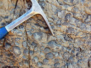
Stromatolites of fossilized oxygen-producing cyanobacteria have been found from 2.8 billion years ago,[10] possibly as old as 3.5 billion years ago.

The biochemical capacity to use water as the source for electrons in photosynthesis evolved once, in a common ancestor of extant cyanobacteria. The geologic record indicates that this transforming event took place early in our planet's history, at least 2450-2320 million years ago (mya), and probably much earlier. Geobiological interpretation of Archean (>2500 mya) sedimentary rocks remains a challenge; available evidence indicates that life existed 3500 mya, but the question of when oxygenic photosynthesis evolved continues to engender debate and research.
A clear paleontological window on cyanobacterial evolution opened about 2000 mya, revealing an already diverse biota of blue-greens. Cyanobacteria remained principal primary producers throughout the Proterozoic (2500-543 mya), in part because the redox structure of the oceans favored photoautotrophs capable of nitrogen fixation.
The most common cyanobacterial structures in the fossil record are the mound-producing stromatolites and related oncolites. Indeed, these fossil colonies are so common that paleobiology, micropaleontology and paleobotany cite the Pre-Cambrian and Cambrian period as an "age of stromatolites" and an "age of algae."
Green algae joined the blue-greens as major primary producers on continental shelves near the end of the Proterozoic, but only with the Mesozoic era (251-65 mya) radiations of dinoflagellates, coccolithophorids, and diatoms did primary production in marine shelf waters take modern form.
Today, the blue-green bacteria remain critical to marine ecosystems as primary producers in oceanic gyres, as agents of biological nitrogen fixation, and -- in modified form -- as the plastids of marine algae.[11]
Classification
The cyanobacteria were traditionally classified by morphology into five sections, referred to by the numerals I-V. The first three - Chroococcales, Pleurocapsales, and Oscillatoriales - are not supported by phylogenetic studies. However, the latter two - Nostocales and Stigonematales - are monophyletic, and make up the heterocystous cyanobacteria. The members of Chroococales are unicellular and usually aggregate in colonies. The classic taxonomic criterion has been the cell morphology and the plane of cell division. In Pleurocapsales, the cells have the ability to form internal spores (baeocytes). The rest of the sections include filamentous species. In Oscillatoriales, the cells are uniseriately arranged and do not form specialized cells (akinetes and heterocysts). In Nostocales and Stigonematales the cells have the ability to develop heterocysts in certain conditions. Stigonematales, unlike Nostocales, includes species with truly branched trichomes. Most taxa included in the phylum or division Cyanobacteria have not yet been validly published under the Bacteriological Code. Except:
- The classes Chroobacteria, Hormogoneae and Gloeobacteria
- The orders Chroococcales, Gloeobacterales, Nostocales, Oscillatoriales, Pleurocapsales and Stigonematales
- The families Prochloraceae and Prochlorotrichaceae
- The genera Halospirulina, Planktothricoides, Prochlorococcus, Prochloron, Prochlorothrix.
Biotechnology and applications
The unicellular cyanobacterium Synechocystis sp. PCC6803 was the third prokaryote and first photosynthetic organism whose genome was completely sequenced.[12] It continues to be an important model organism.[13] The smallest genomes have been found in Prochlorococcus spp. (1.7 Mb)[14][15] and the largest in Nostoc punctiforme (9 Mb)[16]. Those of Calothrix spp. are estimated at 12-15 Mb,[17] as large as yeast.
Some cyanobacteria are sold as food, notably Aphanizomenon flos-aquae and Arthrospira platensis (Spirulina).[18]
Recent Researches have also hinted at their possible application to the generation of Clean and Green Energy via converting sunlight directly into electricity. [19] [20]
Health risks
Certain cyanobacteria produce cyanotoxins including anatoxin-a, anatoxin-as, aplysiatoxin, cylindrospermopsin, domoic acid, microcystin LR, nodularin R (from Nodularia), or saxitoxin. Sometimes a mass-reproduction of cyanobacteria results in algal blooms.
These toxins can be neurotoxins, hepatotoxins, cytotoxins, and endotoxins, and can be toxic and dangerous to humans and animals. Several cases of human poisoning have been documented but a lack of knowledge prevents an accurate assessment of the risks.[21] Recent studies suggest that significant exposure to high levels of cyanobacteria causes amyotrophic lateral sclerosis (ALS). The Lake Mascoma ALS cluster [22] and Gulf War veteran's cluster are two notable examples.[23][24][25]
See also
- Anatoxin
- Archean Eon of earth's prehistory
- Cyanobiont
- Hypolith
- Geological history of oxygen
- The Great Oxygenation Event
- Proterozoic Eon of earth's prehistory
References
- ↑ Ahoren Oren (2004). "A proposal for further integration of the cyanobacteria under the Bacteriological Code". Int. J. Syst. Evol. Microbiol. 54 (Pt 5): 1895–1902. doi:10.1099/ijs.0.03008-0. PMID 15388760.
- ↑ Photosynthetic viruses keep world's oxygen levels up New Scientist 30 August 2009 by Nora Schultz
- ↑ United Nations Conference on Trade and Development,
- ↑ J. Sun, et al. (2004). "Is autoinducer-2 a universal signal for interspecies communication? A comparative genomic and phylogenetic analysis of the synthesis and signal transduction pathways". BMC Evol. Biol. 4: 36. doi:10.1186/1471-2148-4-36. PMID 15456522.
- ↑ E. Dittmann, et al. (2001). "Altered expression of two light-dependent genes in a microcystin-lacking mutant of Microcystis aeruginosa PCC7806". Microbiology 147 (Pt 11): 3113–3119. PMID 11700361.
- ↑ Steve Nadis, The Cells That Rule the Seas, Scientific American, Nov. 2003 [1]
- ↑ Pisciotta JM, Zou Y, Baskakov IV (2010). "Light-Dependent Electrogenic Activity of Cyanobacteria". PLoS One 5 (5): e10821. doi:10.1371/journal.pone.0010821. PMID 20520829.
- ↑ Enrique Flores AH (2008). The Cyanobacteria: Molecular Biology, Genomics and Evolution. Horizon. pp. 3. ISBN 1904455158. http://books.google.com/?id=xgMahO1BXrQC&pg=PA1.
- ↑ Summarised in Cavalier-Smith (2000), Trends Plant Sci, 5: 174-182.
- ↑ Olson JM (2006). "Photosynthesis in the Archean era". Photosyn. Res. 88 (2): 109–17. doi:10.1007/s11120-006-9040-5. PMID 16453059.
- ↑ Herrero A and Flores E (editor). (2008). The Cyanobacteria: Molecular Biology, Genomics and Evolution (1st ed.). Caister Academic Press. ISBN 978-1-904455-15-8 . ISBN 1904455158. http://www.horizonpress.com/cyan.
- ↑ T. Kaneko, et al. (1996). "Kaneko, T. et al. (1996) Sequence analysis of the genome of the unicellular cyanobacterium Synechocystis sp. strain PCC6803. II. Sequence determination of the entire genome and assignment of potential protein-coding regions". DNA Res. 3 (3): 109–136. doi:10.1093/dnares/3.3.109. PMID 8905231.
- ↑ Tabei Y, Okada K, Tsuzuki M (2007). "Sll1330 controls the expression of glycolytic genes in Synechocystis sp. PCC 6803". Biochem. Biophys. Res. Commun. 355 (4): 1045–50. doi:10.1016/j.bbrc.2007.02.065. PMID 17331473.
- ↑ G. Rocap, et al. (2003). "Genome divergence in two Prochlorococcus ecotypes reflects oceanic niche differentiation". Nature 424 (6952): 1042–1047. doi:10.1038/nature01947. PMID 12917642.
- ↑ A. Dufresne, et al. (2003). "Genome sequence of the cyanobacterium Prochlorococcus marinus SS120, a nearly minimal oxyphototrophic genome.". Proc. Natl Acad. Sci. USA 100 (17): 10020–10025. doi:10.1073/pnas.1733211100. PMID 12917486.
- ↑ J.C. Meeks, et al. (2001). "An overview of the genome of Nostoc punctiforme, a multicellular, symbiotic cyanobacterium". Photosynth. Res. 70 (1): 85–106 A number of important advances have occurred in cyanobacterial biotechnology in the recent years. World wide attention is drawn towards cyanobacteria for their possible use in mariculture, food, feed, fuel, fertilizer, colourant, production of various secondary metabolites including vitamins, toxins, enzymes, pharmaceuticals, pharmacological probes and pollution abatement. Only a few cyanobacterial strains (including Spirulina) have been well-characterized or exploited commercially (Thajuddin and Subramanian. Cyanobacterial biodiversity and potential applications in biotechnology. CURRENT SCIENCE, VOL. 89, NO. 1, 10 JULY 2005). doi:10.1023/A:1013840025518. PMID 16228364.
- ↑ M. Herdman, et al. (1979). "Genome size of cyanobacteria". J. Gen. Microbiol. 111: 73–85.
- ↑ Spolaore P, Joannis-Cassan C, Duran E, Isambert A (2006). "Commercial applications of microalgae". J. Biosci. Bioeng. 101 (2): 87–96. doi:10.1263/jbb.101.87. PMID 16569602. http://www.jstage.jst.go.jp/article/jbb/101/2/101_87/_article.
- ↑ Pisciotta JM, Zou Y, Baskakov IV (2010). "Light-Dependent Electrogenic Activity of Cyanobacteria". PLoS One 5 (5): e10821. doi:10.1371/journal.pone.0010821. PMID 20520829.
- ↑ {{|url=http://beta.thehindu.com/sci-tech/science/article477049.ece |doi=10.1263/jbb.101.87}}
- ↑ [2]
- ↑ http://www.als.net/forum/default.aspx?g=posts&t=47305
- ↑ cmd=Retrieve&db=PubMed&list_uids=8830224&dopt=Abstract Cyanobacteria, their toxins and health risks
- ↑ Blue-Green Algae (Cyanobacteria) and their Toxins
- ↑ Harmful Bloom in Lake Atitlán, Guatemala from NASA Earth Observatory, retrieved on 9 January 2010.
Further reading
- Gillian Cribbs (1997) Nature's Superfood, the Blue-Green Algae Revolution. Newleaf. ISBN 0-7522-0569-2
- Marshall Savage, (1992, 1994) The Millennial Project: Colonizing the Galaxy in Eight Easy Steps. Little, Brown. ISBN 0-316-77163-5
- Fogg, G.E., Stewart, W.D.P., Fay, P.and Walsby, A.E. 1973. The Blue-green Algae. Academic Press, London and New York. ISBN 0-12-261650-2
- "Architects of the earth's atmosphere." Introduction to the Cyanobacteria. University of California, Berkeley. 03 Feb. 2006.
- Whitton, B.A. Phylum Cyanophyta (Cyanobacteria). in The Freshwater Algal Flora of the British Isles. Cambridge University Press, Cambridge ISBN 0 521 77051 3
External links
- What Are Cyanobacteria And What Are Its Types?
- Overview of cyanobacteria
- Webserver for Cyanobacteria Research
- CyanoBase
|
|||||||||||||||||||||||||||||||||||
|
||||||||||||||||||||||||||||||||||||||||||||||||||||||||||||||

